Pediatricians, orthopedic specialists, and orthotists all rely on X-ray imaging for accurate diagnosis and ongoing monitoring of spinal curves. It all starts with the initial evaluation of a curve and continues with each new brace fitting which requires updated imaging to ensure proper design and effectiveness. Understandably, many parents have concerns about the cumulative effects of radiation exposure from this frequent imaging.
To help minimize radiation exposure while obtaining high-quality images, National Scoliosis Center offers EOS Low-Dose X-ray Imaging at our Fairfax, Virginia office. When compared to traditional X-ray systems, EOS technology provides detailed, full-body images using approximately one-third of the radiation. This is particularly important for growing children who require multiple scans annually.
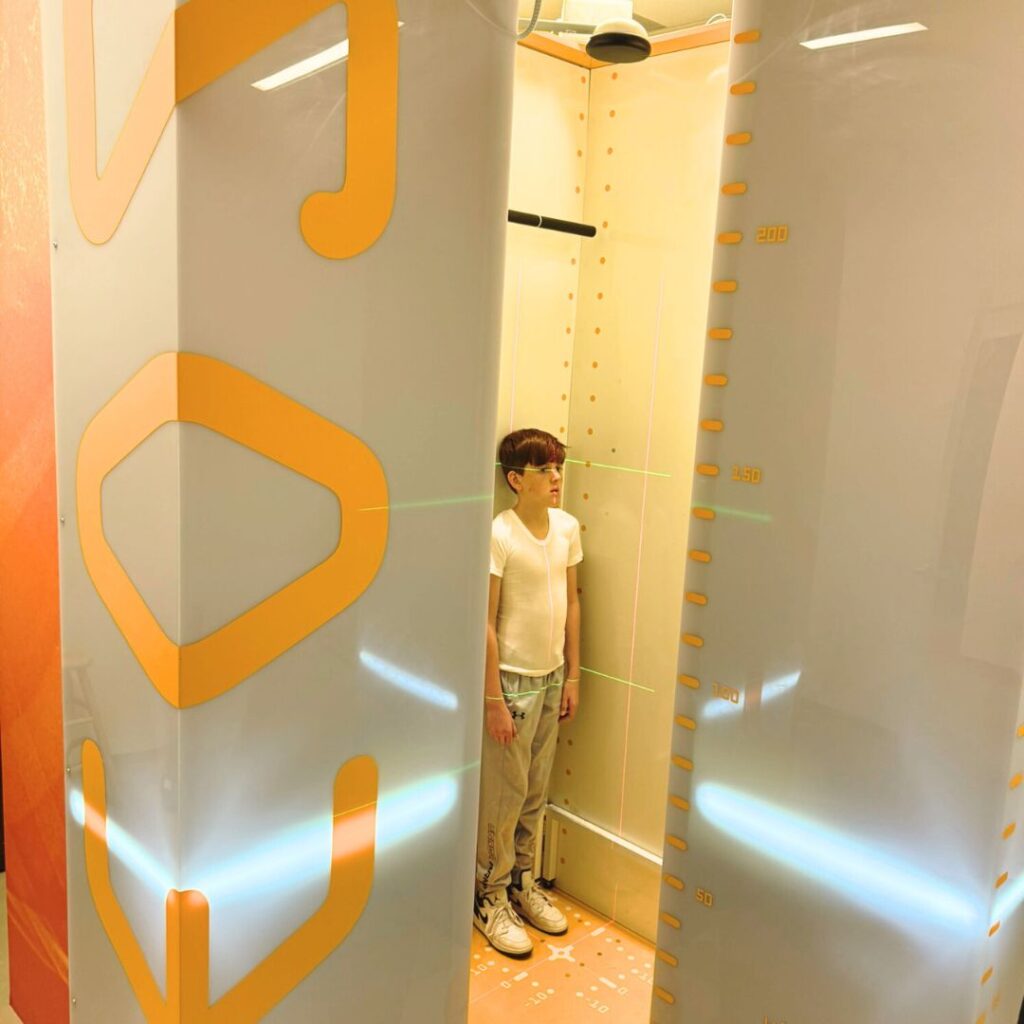
How Does EOS Imaging Work?
Unlike traditional X-rays, EOS captures both front and side views in one scan in about 20 seconds, offering a clear image of the spine and lower limbs without the need for repeated exposures. This full-body view is particularly beneficial in scoliosis treatment, as it allows clinicians to assess spinal alignment and posture in a natural, weight-bearing position.
Another advantage of EOS is that the imaging process is more comfortable for patients, allowing them to stand or sit upright while the two simultaneous X-ray beams scan the body. This approach reduces the need for awkward positioning, decreases overall scan time, and makes the experience easier for children and adolescents.
Why Low-Dose Imaging Matters
Minimizing radiation exposure is a priority for all clinicians treating scoliosis as well as those families with a child diagnosed with scoliosis.
At National Scoliosis Center, we are committed to utilizing advanced, research-supported technologies that prioritize both safety and effectiveness in scoliosis treatment. Our EOS imaging system ensures ultra-low-dose radiation while providing patient-friendly, high-quality images for precise scoliosis assessment, brace design, and the ongoing monitoring of spinal curves.